The salad is an undeservedly neglected part of holiday lunch. Hungarians just eat something pickled with the meat and call it a day, while food bloggers throw together something from the leftovers. Yet there’s real science in salad, let me tell you! The word “salad” comes from the Latin expression herba salata (meaning “salted herb”). So I ask you, why is it that we salt cucumbers—and quite a few other vegetables—when making salad? And why don’t we do the same with, say, lettuce?
According to Hungarian writer Gyula Krúdi, cucumber salad is “a dish for which the skill of cooks alone is not enough; it also requires complete love of humanity, a deep understanding of appetite, a sense of the proper mood, and the ability to capture the right disposition.” One thing is certain: it’s not easy to make a cucumber salad that will fully satisfy a true gourmand.
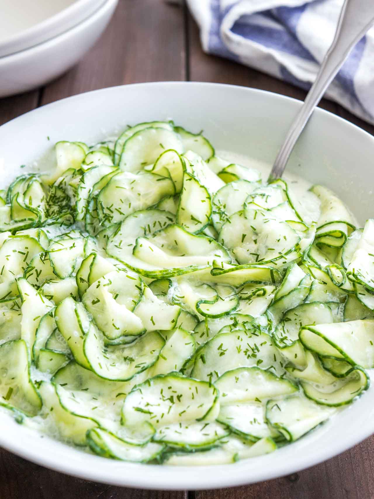
The traditional recipe
The traditional recipe calls for slicing the cucumber into thin rounds, salting it lightly, and letting it sit for about half an hour. Then it’s squeezed, and a salt-free, vinegar-based dressing is poured over it. This dressing typically contains fresh herbs, white pepper, garlic, paprika, sugar, lemon juice, and olive oil to taste. The dish becomes truly delicious when served with sour cream or yogurt on top.
But why do we salt the cucumber at the beginning if we’re just going to squeeze out the salty liquid afterward? And do we pour valuable vitamins and minerals down the drain along with that liquid?

Required Equipment
- A red onion (for the epidermis sample)
- Scalpel or a sharp knife (to cut a small piece)
- Tweezers (for peeling off the epidermis)
- Microscope slide (glass slide)
- Cover slip
- Dropper (to place a drop of water on the slide)
- Tap water
- Light microscope (with at least 100× magnification, ideally 400×)
Experiment with Red Onion
To find the answer, we first need to observe what actually happens during salting. We’ll conduct the experiment not with cucumber, but with red onion, because the effect is easier to see on onion, and it makes no difference to the principle whether we salt onion or cucumber.
Cut out a small piece from one of the fleshy inner layers of the onion, and peel off the purple epidermis—the thin outer membrane. It doesn’t matter if it doesn’t come off perfectly; what we need is a thin section, ideally one cell layer thick, at most two, so it’s easy to examine.
Place a drop of tap water on a clean microscope slide. Drop the peeled layer into the water, gently spread it out on the slide, then cover it with a cover slip and place it under the microscope.

Under the microscope, you can see elongated cells closely packed together. These are the epidermal cells of the onion. Two cell structures are clearly distinguishable: the cell wall, made primarily of cellulose, and the vacuolar system, which appears purple due to anthocyanin pigments.
The vacuoles are fluid-filled compartments enclosed by a membrane, containing not only pigments but also various dissolved salts and organic acids. Under normal conditions, the vacuoles almost completely fill the cells. The colorless cytoplasm appears only as a thin, tubular layer along the cell wall.
If your microscope is good and you observe very carefully, you might also spot a few rounded, colorless nuclei, but these are barely visible because their refractive index is nearly the same as that of the surrounding cytoplasm.

When you observe the onion peel under the microscope, you’ll see a neat arrangement of elongated, brick-like cells pressed closely together. These are epidermal cells forming the onion’s protective tissue.
Key features you can notice:
- Cell wall: Thick, transparent structure mainly composed of cellulose, providing shape and rigidity.
- Vacuole: The largest internal compartment, filled with cell sap. It contains water, dissolved salts, organic acids, and anthocyanin pigments, which give the purplish-red color.
- Cytoplasm: Appears as a very thin layer lining the inside of the cell wall, since the vacuole occupies most of the cell’s volume.
- Nucleus: Usually round and colorless, hard to see because it blends with the cytoplasm.
Under normal conditions, the vacuoles almost completely fill the cells, so they look plump and tight. This state will change dramatically when salt is introduced—a key step in our cucumber salad story.
If we were to stain the peel (by treating it with methylene blue or blue ink for 1–2 minutes, then rinsing it with tap water), the nuclei would become clearly visible. If we want to observe the stained specimen for an extended period, we should cover it not with plain tap water but with a 1:1 mixture of water and glycerin.

Apart from the nucleus, there are other organelles in the cell, but none of these are visible under a light microscope. They would only appear under an electron microscope—but that’s fine, because right now we’re only interested in the vacuoles. We chose red onion because anthocyanins color its vacuoles purple. Anthocyanins are natural pigments found in many things—from red wine to raspberries to red cabbage. Their molecules are fairly large and cannot pass through the membrane surrounding the vacuole.
Once we’ve had our fill of admiring the view, it’s time to salt the onion. Prepare a concentrated solution from table salt and a little water. Place a generous drop of this solution on the microscope slide at the edge of the cover slip, and on the opposite edge touch a strip of blotting paper. As the blotting paper starts drawing water out from under the cover slip, the salt solution will flow in from the other side and gradually reach the onion.

Looking through the microscope, we observe that the vacuoles (and, in fact, the entire cell including the cytoplasm) detach from the cell walls, shrink within 4–5 minutes, and their color becomes darker.
The explanation for this phenomenon is that the salt concentration on both sides of the cell membranes tends to equalize. Normally, there is some dissolved salt in the cytoplasm and in the extracellular space as well, and these are in balance. When we introduced the salt solution to the onion, this balance was disrupted—the salt concentration outside the cell became higher than that of the cell sap.
The dissolved salt ions cannot diffuse through the membranes, but water can. The balance between the outer hypertonic solution (with higher salt concentration) and the cytoplasm can only be restored if water flows out of the cell into the external space through the cell membrane. As the cell loses water, the cytoplasm and the vacuoles inside it shrink accordingly.

The same amount of salt and pigment remains in less water, which means their concentration increases. We cannot see the salts, but we can observe the pigment becoming more concentrated: the vacuoles appear darker. For this phenomenon, only the membranes matter—the elongated, honeycomb-like network formed by the cell walls does not impede the movement of fluids and dissolved substances.
However, the rigid cell wall cannot follow the shrinking process, so the plasma enclosed by the cell membrane gradually pulls away from the wall, and plasmolysis occurs. By the end of the process, the vacuoles have contracted into smaller or larger spherical shapes in the center of the space inside the cell wall, and around them the transparent cytoplasm is now more visible. Under higher magnification, you can also see thin strands of cytoplasm stretching from the contracted plasma toward neighboring cells.
Plasmolysis is a reversible process. If we now use the same method to introduce pure distilled water under the cover slip, the vacuoles will start to expand. The distilled water dilutes the solution between the cells, reducing the salt concentration in the external environment. The balance is restored by water flowing back into the cells, which begin to swell. In a hypotonic solution—where the salt concentration is lower than in the normal cell sap—the cells swell beyond their normal size because they can only reduce the higher internal salt concentration by taking in water. They outgrow the space provided by the cellulose cell walls, bulge out, and eventually burst and collapse.

Plasmolyzed Cell (after salt treatment)
- Cell wall remains in place, forming the rigid outline.
- Inside, the cytoplasm and vacuoles are shrunken and detached from the wall, appearing as a smaller, darker mass in the center.
- Label: Plasmolyzed state – Water leaves the cell in hypertonic salt solution.
Restored (after adding distilled water)
- Vacuoles have expanded again, filling the cell’s interior.
- Cytoplasm returns to its original position, pressing against the cell wall.
- Label: Deplasmolysis – Water re-enters the cell in hypotonic solution.
Those who believe in the healing power of distilled water should think carefully about this experiment. In medical practice, it is precisely to avoid the phenomenon we just demonstrated that isotonic, so-called physiological saline is used instead of pure water. This 0.9% salt solution is in osmotic balance with human tissue cells, meaning their volume does not change when such a solution is applied to them.
What Happens in Cucumber Salad?
The onion peel experiment showed that salting causes the cells to shrink. Cucumbers have a very high water content—around 95%, like most gourds. When salted, they lose some of this water. After the salty liquid is removed, the cook pours slightly sweetened water with vinegar or lemon juice over the cucumber slices. Vinegar and citric acid (and generally weak acids that are poorly dissociated) can pass through cell membranes more easily. As a result, the cucumber absorbs a good amount of vinegar, giving us a much more flavorful salad than if we simply poured the dressing over it.
The purpose of salting, then, is dehydration—removing water, which is then replaced by a tastier solution.
In our plasmolysis observation, we saw that the purple pigment does not escape into the extracellular space because it cannot pass through the membranes. Aside from water and the acids mentioned, only small, nonpolar molecules (e.g., ethanol, oxygen, carbon dioxide, etc.) can cross the membrane. Dissolved salt ions, larger polar molecules (such as glucose), and macromolecules (such as proteins) cannot. When cucumbers are salted, water leaves the cells, but valuable minerals and vitamins remain.
In fact, salting (and sugaring, which similarly reduces water content) is a very old food preservation method, the essence of which is to deprive spoilage-causing microbes of the water they need to grow. Laboratory tests show that this process does not harm the nutritional or vitamin content of foods—in fact, quite the opposite: because of water loss, the food becomes more flavorful and relatively richer in vitamins.
The downside is that the salt (or sugar) content of the food increases. In salted fish, salt has been shown to cause some breakdown of vitamin B1. This likely happens in cucumbers as well, but overall, salting does not harm cucumber salad—in fact, it makes it better.
So why don’t we salt lettuce, for example? The reason is probably primarily aesthetic and related to texture. Due to plasmolysis caused by salting, turgor pressure (the internal fluid pressure against cell walls) decreases, which outwardly appears as limp, floppy leaves—like a deflated air mattress. Such salad leaves don’t look very appetizing. However, if we just add a little salt to the dressing and don’t let the leaves sit in it for too long, we get a fresh, crisp, and flavorful salad.

An Experiment with a Mega Cell
Even those without a microscope can observe cell size changes in a salt solution. All you need is a cell large enough to see with the naked eye. That cell is the chicken egg, which, although an animal cell, has membranes that behave similarly to plant cell membranes.
To perform the experiment, we need to remove the eggshell. Place the egg in a glass and pour in just enough household hydrochloric acid (HCl) so that it does not completely cover the egg. The acid will start dissolving the shell with vigorous fizzing. Be careful—the caustic foam that forms can overflow the glass! To avoid accidents, wear safety goggles and rubber gloves.
After a few minutes in the acid, the shell dissolves, leaving only the elastic membrane, through which the yolk is faintly visible. Carefully pour off the acid, rinse the egg, and cover it with water. By the next day, the egg will have noticeably swollen from absorbing water. The reason is the same as plant cells swelling during deplasmolysis.
For children, use vinegar instead of acid! Vinegar will also remove the shell overnight, but it is far less dangerous than hydrochloric acid. An egg soaked in vinegar will absorb the vinegar-water in 2–3 days. If you pierce it with a needle, water will squirt out. If you then place the swollen egg in concentrated salt solution, it will lose water and shrink—just like the onion cells.